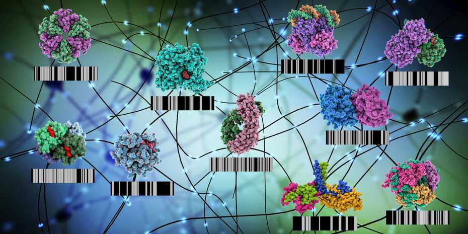
ETH researchers have drastically improved existing proteomics techniques so they can capture all functional alterations in proteins. Their work paves the way for using these signatures as diagnostic tools.


In biological cells, proteins are everywhere: these building blocks of life perform countless important functions. A human cell contains thousands of different proteins at any given time, often with copies of each protein type present in their hundreds or even thousands simultaneously.
In recent years, researchers have succeeded in using measuring instruments to capture this enormous diversity in full. Today, it’s possible to screen cells, organs or even whole organisms in one go to record their entire proteome, in other words all the various protein species and their quantities. Traditional proteomics techniques can also determine how the abundance of each protein species changes in response to changing environmental conditions.
Screening failed to detect many functional changes
Until now, though, standard proteome screening was unable to detect simultaneously many of the molecular events that occur in cells and lead to changes in protein function.
These events include chemical changes to the proteins themselves, such as phosphorylation, as well as interactions with other proteins or molecules. Such molecular events are important: many processes in cells, such as signalling cascades, often rely exclusively on various molecular events rather than on adjustments to protein levels. This allows a cell to adapt very quickly to new circumstances with its existing set of protein molecules, rather than having to make new ones.
Making functional changes measurable
This led Paola Picotti, Professor of Molecular Systems Biology at ETH Zurich, and her team of researchers to suspect that structural changes could be used as a readout (or a “proxy”) for all the various molecular events that result in protein functional changes.
The systems biologists refined an existing approach for mapping proteomes so they could detect all such functional changes in proteins simultaneously down to the last detail.
They have now succeeded in this endeavour, as they recently reported in the journal Cell. Their new approach lets the researchers measure many enzyme activities molecular interactions and chemical modifications in situ, i.e. directly in cell fluids.
Coverage massively increased
To use them as a proxy for molecular events, the researchers measure the protein structures present in a sample. They measure their quantities at the same time. “This way, we capture the majority of events that affect protein function – a dramatic increase in coverage,” Picotti says.
“We can now detect a much higher number of altered biological processes than by measuring protein abundances alone”
-Paola Picotti
The ETH professor laid the foundation for this new method a few years ago. Using a technique called limited proteolysis mass spectrometry (LiP-MS), she and her colleagues were able to relatively easily probe the structures of a vast number of proteins in a biological sample, such as yeast cytoplasm or body fluids from biopsies. This also enabled her to record various structures of one and the same protein.
Making proteins into better biomarkers
To test the new LiP-MS approach, Picotti and her colleagues took a close look at three well-established systems. They found that the method not only captured all known functional changes, but also uncovered previously unknown events. “That means we can now detect a much higher number of altered biological processes than by measuring protein abundances alone,” Picotti says. This is an important step towards making proteins, their altered structures and their functions more usable as valid biomarkers in the future.
One way the researchers tested their concept was on yeast cells, by subjecting them to salt stress. After a short time, the cells set in motion a special pathway to cope with the increased salt concentration in their environment. When this signalling pathway is activated, some proteins become phosphorylated – they have phosphates “attached” to them – while some others interact with additional molecules, and yet others change their activity. This caused the enzymes to change their structure – and thus also their function.
The ETH researchers found that among the total of 3,500 different protein types present in yeast, more than 300 changed shape in this experiment, while only 30 of them changed in abundance. “This means cells don’t produce particularly large numbers of proteins in response to short-lived new stimuli or acute stress; rather, they remodel existing ones and modify their function,” Picotti says.
Once the stress has been overcome, the reshaped molecules can be restored to their original state. As a result, the number of proteins in cells remains fairly constant over time. This is good news for cells, since producing new molecules takes a lot of time and energy. In contrast, reshaping them takes just a few seconds and little energy.
Promising diagnostic tool
Picotti has already conducted a project to test the method on a human disease. She and her colleagues compared several hundred samples from Parkinson’s patients with those from healthy people to find structural biomarkers for the disease.
Picotti’s preliminary conclusion is that “the quantity of proteins alone is not diagnostically useful in this case,” with the majority of proteins approximately equally abundant in both healthy and diseased individuals. However, when the researchers looked for different structures of the same proteins, things appeared more promising. Numerous proteins showed altered structures in the samples taken from patients, which suggests that the approach has promise as a diagnostic tool. Whether it might also serve as a means of early detection, however, remains to be explored.
ETH spin-off offers method commercially
The researchers have already patented their approach. The ETH spin-off Biognosys, which specialises in proteomics, has licensed the method and is successfully offering it as a commercial service to analyse drug development samples for structural changes in proteins. The new approach is also attracting a great deal of interest in the academic world. “I get several requests a week from researchers abroad to see if my lab can analyse samples for them. But we can’t fulfil all these requests because we don’t have the capacity,” Picotti says.


