An estimated 19.3 million new cancer cases and almost 10 million cancer deaths occurred worldwide in 2020.


It’s estimated that one in three individuals will be diagnosed with some kind of cancer in their lifetime. When cancer is diagnosed at a stage where it’s not responsive to treatments, it usually becomes a fatal disease.
It’s for this reason that the race to identify tools that enable early cancer detection is vital.
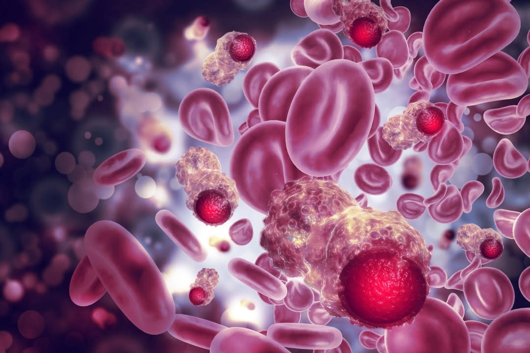
Cells can communicate with each other. They communicate with neighbouring or distant cells by secreting lipid-bound vesicles known as extracellular vesicles (EVs). All types of cells discharge EVs, which provide information about the originating cells and their physiological state.
Because EVs can give these pieces of information, particularly in cancer progression, they’ve emerged as a novel source of non-invasive cancer biomarkers that could help diagnose and treat cancer early.
One of the reasons why cancer causes so many deaths is because of late diagnosis. Diagnosing cancer ahead, even before clinical symptoms appear, would significantly increase the survival rate of patients.
But, an early diagnosis has proven to be challenging, as imaging techniques often detect tumour mass only when it’s visible –which is at a later stage.


EVs contain some cancer-specific proteins involved in diverse processes such as proliferation and malignancy, which are useful for cancer to spread, thus making them useful as biomarkers. Examining EVs can increase the likelihood of early cancer detection, as it allows rapid, inexpensive, non-invasive, and comparatively accurate diagnosis.
Still plenty to learn
However, there’s still much to learn about EVs and their association with cancer.
“My research work advances the understanding of how EVs help cancer progression by promoting cancer cells’ survival and mediating drug resistance,” says Dr Lee Wai Leng, from the School of Science at Monash University Malaysia.
The role of EVs in mediating drug resistance was confirmed when Dr Lee and her team inhibited EV secretion, decreasing the viability of the cells. This suggests that inhibition of EV release could be a novel therapeutic approach to sensitise drug-resistant cells to chemotherapy.
To date, drug resistance remains a critical obstacle in most chemotherapeutic treatments.


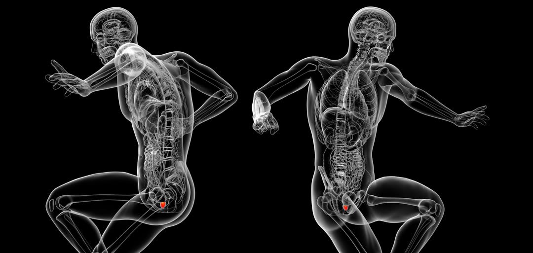
Dr Lee and her team demonstrated that it was possible to detect cancer-specific proteins by examining the urine of patients with prostate cancer against healthy participants. The team analysed the EVs’ composition using a Fourier-transform infrared (FTIR) spectroscopy, which helped them accurately determine which EVs originated from prostate cancer patients and healthy participants.
“The data suggests we could use this technique for the early screening of cancer, and enable the accurate staging and grading of the disease,” Dr Lee added.
Detecting EVs could also help monitor the response to treatment, and the possibility of a relapse.
EVs as theragnostic markers
In one of their studies, Dr Lee and her team discovered several EV proteins that can potentially be developed as theragnostic markers (biomarkers that can be used for both therapy and prognosis), indicating how responsive patients are to cancer treatment.
This finding is essential for the development of personalised medicine, also called precision medicine, aiming to tailor therapy with the best response and highest safety margin to ensure better patient care.
Besides testing urine samples from prostate cancer patients, the team plans to test the efficacy of different body fluid types from other types of cancer.
The team’s “UEV diagnostic: Non-invasive test for prostate cancer detection” won a silver medal at the 31st International Invention, Innovation and Technology Exhibition (ITEX) 2020, Malaysia.
The stakeholders involved in this research are government agencies and life science industries interested in developing EV-based technology for disease detection or treatment.


