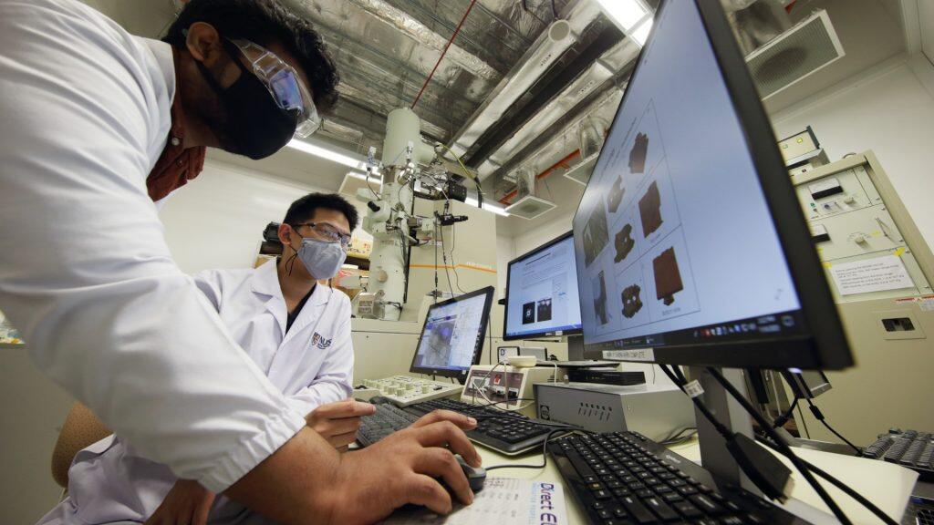

Researchers at the NUS Centre for BioImaging Sciences (CBIS) have developed a new method to observe materials of low atomic weight, such as biological molecules, under an electron microscope, while minimising damage to the sample. Assistant Professor Duane Loh and Research Fellow Dr Deepan Balakrishnan accomplished this by dramatically reducing the intensity of the microscope beam, and using computer analysis to reconstruct the image from the weak signal.
Of the six electron microscopes at CBIS, the one used by the duo is unpopular with other researchers for atomic-resolution imaging because its sample holder is more prone to random mechanical movement. For a transmission electron microscope with a resolution of 0.1 angstrom, or a hundred millionth of a millimeter, there is a lot of motion blur if it is not dampened properly. But it is perfect for testing their algorithm.
The two of them tried their technique on a thin layer of tiny gold nanocrystals, each just 10 to 20 atoms across. Using a data compression method newly developed by Dr Abhik Datta, another researcher in the lab, they rapidly extracted the faint electron signals as they trickled in over the course of an hour, then they fed these signals into the computer. Not long after, the gold nanocrystals magically appeared on screen, sharp and clear. This was achieved using a statistical learning model based on the physics of how the microscope forms a noisy image. The computer corrects for the motion blur by realigning the electron signals to recover the most statistically likely shape of the nanocrystal.
The technique allows imaging at far lower dose rates and longer exposure times than was previously possible, and is useful for examining biological molecules or anything else containing lighter elements which are prone to damage by the electron beam.
This study was funded by the Competitive Research Programme under Singapore’s National Research Foundation.Using machine learning to recover a sharp image of moving gold nanoparticles (far right) from very sparse electron hits (far left) accumulated over time. (Image: Dr Deepan Balakrishnan)


Fusing disciplines for scientific research
Asst Prof Loh’s research in what he calls “computational lenses” exemplifies a lot of what goes on at CBIS – melding traditional microscopes with computer science to solve problems in new ways.
The equipment at CBIS is not just about making nice images of golden nanocrystals. The biological imaging and computational technologies there serve as the backbone of many multidisciplinary and translational research projects conducted at NUS. For example, to design a high-tech material, computational lenses can be used to discover hidden molecular patterns, and how they can be altered to meet different needs. To understand how a complex protein molecule causes cancer, millions of pictures of it under natural conditions can be taken, and computationally reconstruct its 3D shape in unprecedented detail.
These techniques were all developed at CBIS this year, and they complement different research projects that fall into NUS’ key areas of focus, such as in Materials Science, as well as Biomedical Science and Translational Medicine.
Professor Thorsten Wohland, Director of CBIS and Vice-Dean for Research and Space at NUS Science, shared, “Microscopes can see over a much wider wavelength range than humans. They extend our senses and give us a new picture of the world. But now the picture is not enough – we have to analyse the numbers that make up the image.”
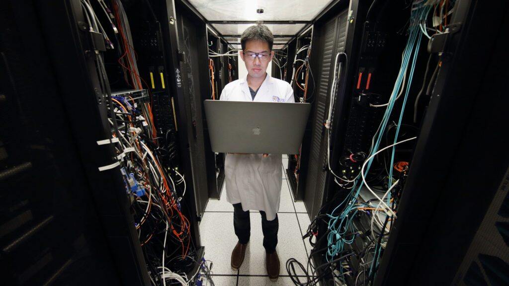
With the advancement in technology, CBIS now not only possesses sophisticated microscopy and imaging techniques that can generate atomic structures and measure dynamics simultaneously and in real time, but also has a strong information technology infrastructure and powerful computational methodologies that are able to interpret and provide the fullest picture yet of biological systems. In fact, CBIS is unusual among microscopy facilities in having its own server to fulfill its computational needs. This combination provides scientists with the means to deepen understanding of complex issues in biology.Assistant Professor Duane Loh working on his computational lens algorithms.

Fueling interdisciplinary research
CBIS is also a melting pot for physicists, chemists, biologists and computer scientists. Asst Prof Loh, for example, holds joint appointments in NUS Physics and NUS Biological Sciences, while serving as one of the principal investigators at CBIS, and infusing his dual disciplines with computer science.
For postdoctoral fellows and graduate students who conduct their research in the Centre, there is a mixed-seating area where they are encouraged to mix across disciplines. This arrangement allows them to bounce ideas off one another, planting seeds that may one day sprout into solutions for problems that have long confounded scientists, or simply to ignite collaborations. And when they need a microscope, CBIS is a paradise.
Besides the six electron microscopes in the Centre, there is also a room filled with seven state-of-the-art optical microscopes. CBIS also is part of SingaScope, Singapore’s national microscopy infrastructure network. The facilities under this network are available to all researchers and industry.
Microscopy has come a long way since the 17th century, when Robert Hooke made drawings of cork cells and Antonie van Leeuwenhoek reported his observations of animalcules at the Philosophical Transactions of the Royal Society. Today, some microscopes are so big that they need their own room, and so powerful that they can see atoms.
And to top it off, they have been bestowed near-magical powers by computers.


